
A Patient’s Guide to Coronary Angiography: Understanding Procedure, Types, Safety & Key Differences
Coronary angiography is an important diagnostic method used to study how well the coronary arteries are functioning. Many individuals learn about this procedure when they experience symptoms that could be linked with the heart, and they want to understand what actually happens inside their arteries. Being informed helps patients feel more prepared, and it supports better discussions with their treating doctor. In many cases, individuals search for clarity from trusted healthcare sources, including the Best Cardiologist in Pune, especially when they want to understand cardiac evaluations or explore options like heart blockage treatment without surgery in Pune. This blog offers a clear, medically accurate, and easy-to-follow explanation for anyone wanting to understand coronary angiography from a patient’s perspective.
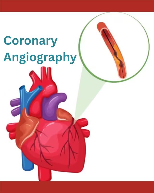
What Is Coronary Angiography?
Types of Angiography
1. Coronary Angiography
Used to visualise the coronary arteries supplying the heart. This is the most commonly performed type.
2. CT Coronary Angiography
A non-invasive imaging technique performed using CT scanning and contrast dye. It helps in evaluating plaque and vessel structure without inserting a catheter.
3. Peripheral Angiography
Used to examine arteries in the legs, arms, or other areas when symptoms suggest reduced blood flow.
4. Cerebral Angiography
Used to visualise blood vessels supplying the brain.
5. Pulmonary Angiography
Used to view arteries in the lungs, particularly when pulmonary embolism is suspected.
Each type has its own purpose, but all follow the principle of using contrast dye and imaging to study blood vessels.
What Happens During the Procedure?
1. Preparation
The patient lies on a table in a cardiac catheterisation laboratory. Heart rate, oxygen levels, and blood pressure are monitored. The insertion site—commonly the wrist or sometimes the groin—is cleaned and numbed with a local anaesthetic.
2. Catheter Insertion
A thin, flexible tube known as a catheter is gently inserted into the artery. Using continuous imaging guidance, the catheter is navigated toward the coronary arteries.
3. Injection of Contrast Dye
Once in position, contrast dye is released through the catheter. This dye makes the arteries visible on X-ray images, allowing the doctor to study the flow of blood and any narrowing present.
4. Imaging
Images are captured from multiple angles to give a clear view of the coronary arteries. This helps identify areas where the vessel walls may be narrowed or where blood flow might be reduced.
5. Completion
After imaging, the catheter is removed. A band or pressure device is placed on the wrist or groin to reduce the risk of bleeding. Patients rest for some time while being monitored.

What Conditions Can Angiography Diagnose?
- Narrowing of the coronary arteries
- Blockages caused by plaque buildup
- Abnormal vessel structure or congenital variations
- Reduced blood flow in certain segments of the heart
- Damage from previous heart events
- Conditions related to chest discomfort or unexplained breathlessness
What Is the Difference Between Angiography and Angioplasty?
- A diagnostic test.
- Used to visualise coronary arteries.
- Involves injecting dye and taking images.
- Helps identify blockages or narrowing.
- A therapeutic procedure.
- Performed if significant narrowing is identified.
- A balloon may be used to widen the vessel.
- A stent may be placed to maintain vessel openness.
Risks and Complications
- Minor bleeding or bruising at the insertion site
- Temporary discomfort during catheter placement
- Rare allergic responses to contrast dye
- Rare irregular heart rhythm during the procedure
- Infection at the insertion site (uncommon)
- Very uncommon risk of vessel-related injury
Many people seek information from trusted sources or healthcare professionals when they are trying to understand diagnostic tests or potential treatment pathways. Reliable explanations support clarity and confidence in decision-making. As patients continue to learn more about coronary evaluations and arterial health, discussions with a qualified doctor or the Best Cardiologist in Pune can help address personal concerns and guide next steps based on individual medical needs.